Neuroimaging
Neuroimaging services from UMass Memorial Health include sophisticated brain scans and other studies that help you receive the best possible care.
Why Choose Us for Neuroimaging?
Uncommon Expertise
Our neuroimaging specialists are radiologists with additional training in brain, spine and nervous system imaging. We offer a depth of experience in all aspects of neuroimaging, including rare and complex conditions. We perform tens of thousands of studies each year and provide a level of expertise that no other program in Central Massachusetts can offer.
Collaborative Care
For complex conditions, neuroradiologists are often part of a multispecialty team. We provide direct input into treatment planning so that you receive the best available therapies. We frequently work alongside experts in neurosurgery, neurology, cancer care, otolaryngology (head and neck surgery), spine surgery and many other providers.
Advanced Interventional Procedures
Some neuroradiologists have additional training in minimally invasive techniques that enable us to deliver treatments without surgery. We use this method to provide advanced care for brain tumors, back pain, stroke and vascular malformations. Get more information about neurointerventional radiology.
Our Radiology Locations
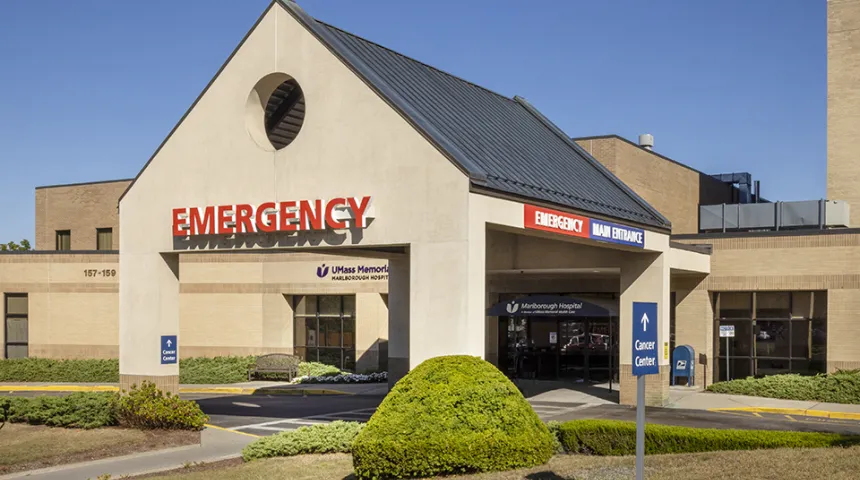
UMass Memorial Medical Center - Marlborough Campus
157 Union Street,
Marlborough, MA 01752

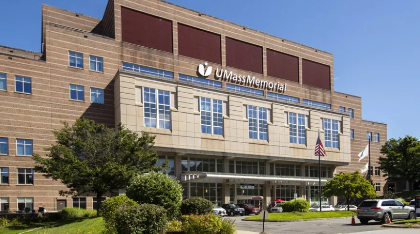
UMass Memorial Medical Center - University Campus
55 Lake Avenue North,
Worcester, MA 01655
UMass Memorial Medical Center - Memorial Campus
119 Belmont Street,
Worcester, MA 01605
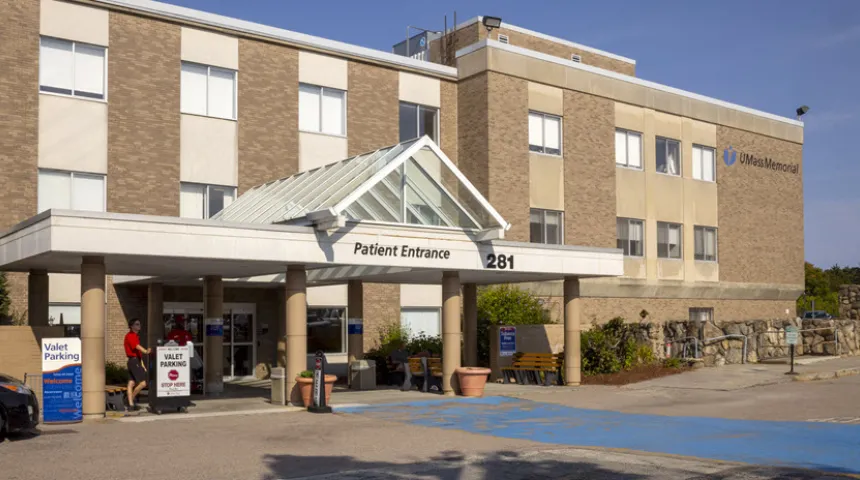
UMass Memorial Medical Center - Hahnemann Campus
281 Lincoln Street,
Worcester, MA 01605
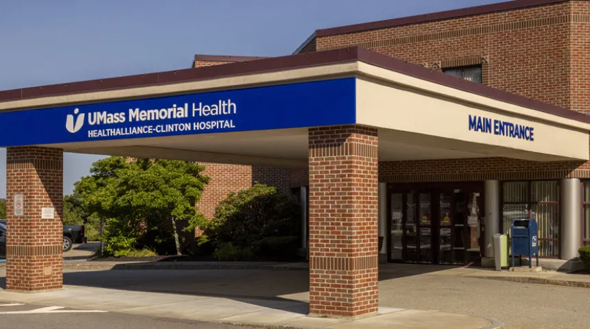
HealthAlliance-Clinton Hospital - Clinton Campus
201 Highland Street,
Clinton, MA 01510
HealthAlliance-Clinton Hospital - Leominster Campus
60 Hospital Road,
Leominster, MA 01453
Refer a Patient
UMass Memorial providers can place orders or refer patients through EPIC. Community providers can receive assistance through Physician Concierge Services (PCS). You can also reach PCS by calling 800-431-5151 or emailing pcs@umassmemorial.org.
Get Started
Patients can call 855-UMASS-MD (855-862-7763) to schedule a neuroimaging study.