Mammography and Breast Imaging
We offer standard and advanced imaging services that detect breast cancer in early stages — well before you notice symptoms.

Why Choose Us for Breast Cancer Screening and Imaging Services?
Expertise
Highly skilled radiologists interpret your breast imaging studies. The team includes specialists with advanced fellowship training in breast imaging, so you can feel confident about the accuracy of your results. Our experience interpreting a high volume of scans lets us quickly determine whether masses are benign or cancerous. The result is timely care for you.
Comprehensive Breast Program
When you choose UMass Memorial for your mammogram, you have access to a nationally-recognized Comprehensive Breast Program. This includes personalized care for high-risk patients, genetic testing and the most advanced and compassionate care when you need it. Learn more about our nationally recognized Breast Cancer Center.
Leadership
We train the next generation of breast imaging experts with a fellowship program offered through our partnership with UMass Chan Medical School. We provide advanced training to radiologists who wish to specialize in breast imaging. Only centers such as ours that perform a high volume of scans with the highest standards maintain this honor.
Our Mammography Locations
Equipment and hours of operation vary by location.
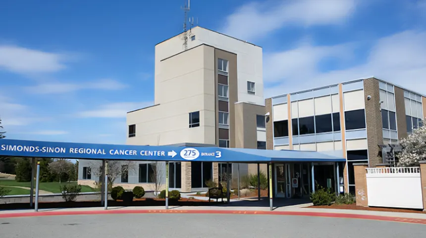
Simonds-Sinon Regional Cancer Center
275 Nichols Road,
Fitchburg, MA 01420
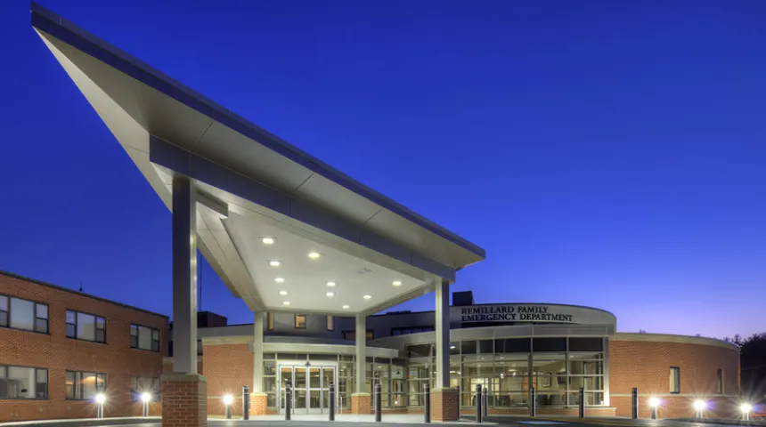
Harrington Hospital - Webster Campus
340 Thompson Road,
Webster, MA 01570
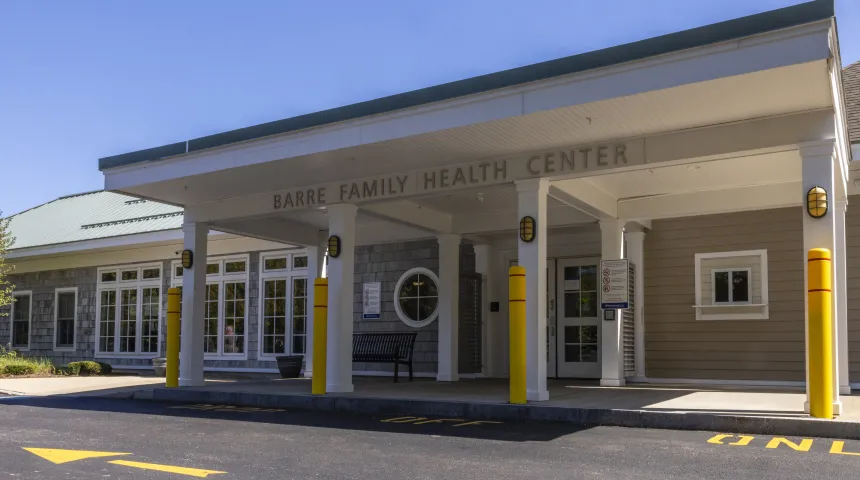
Barre Family Health Center
151 Worcester Road,
Barre, MA 01005

Tri-River Family Health Center
281 East Hartford Avenue,
Uxbridge, MA 01569
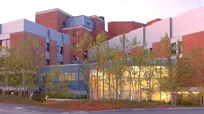
Milford Regional Medical Center
14 Prospect Street,
Milford, MA 01757
Get Started
Call 855-UMASS-MD (855-862-7763) to schedule an appointment.
Schedule a Mammogram Online via myChart
For women over 40 with a primary care provider and myChart account, you can now schedule a best-in-class annual screening 3D mammogram (tomosynthesis) at one of our many convenient locations. Visit myChart and schedule your annual appointment today!
Online scheduling is only available for 3D mammograms, which may exclude certain locations, and for patients who do not already have a scheduled appointment.